Email: info@ambayeyehospital.com Call: +91-0161-4609810 Time: OPD (8:30 AM - 2:00 PM) (5:00 PM - 6:30 PM)

MEL 80 EXCIMER LASER
The MEL 80 is a top quality Carl Zeiss Meditec product, designed to make the correction of vision defects even safer, more patient-friendly and individual. All the parameters of this ultramodern work platform are oriented towards increasing efficiency, achieving optimum treatment results and the rapid recovery of vision. Key factors here are the extremely fast ablation, customized treatment planning with the optional CRS-Master, the high-performance eyetracker system and the “Eye Registration” torsion compensation system.
FEATURES:-
- The exceptionally fast MEL 80’s short ablation time reduces procedure time for greater patient comfort.
- Shortened stroma exposure time means faster visual recovery.
- Very small 0.7 mm spot permits the finest corrections without losing the benefits of smooth ablation.
- Two specially optimized ablation profiles to choose from help you produce excellent results.
- An active eye tracker with excellent feedback times and an ultrarapid IR camera catching both pupil and limbus provides exact positioning during the laser treatment.
ZEISS CRS MASTER
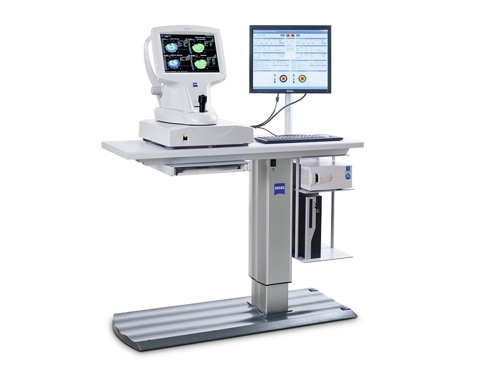
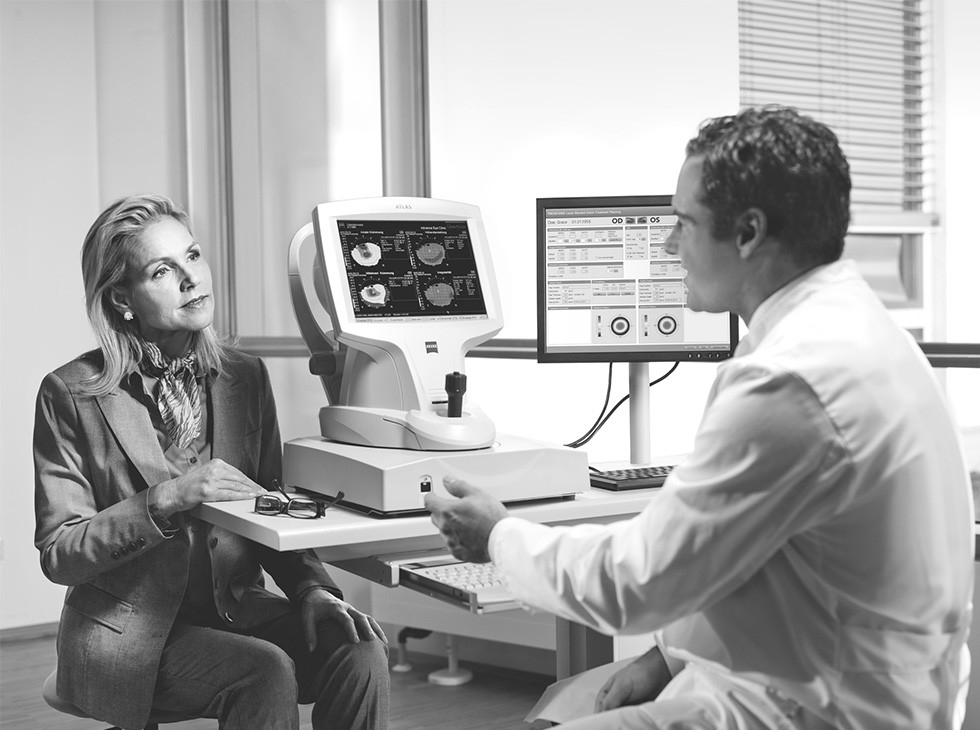
The CRS-Master® is a flexible and efficient remote treatment planning station for conventional and customized laser vision corrections, including LASIK, Femto-LASIK, PRK and LASEK. It also enables binocular treatment planning for presbyopia patients using the unique PRESBYOND® Laser Blended Vision option from ZEISS. An intuitive planning software and an integrated corneal topography system (ATLAS®9000) enables you to merge relevant patient and diagnostic data in just one workstation, creating a complete overview for a treatment planning considering all relevant information.
With the CRS-Master from ZEISS, treatment planning is greatly simplified. The versatile planning station conveniently combines all relevant measurement and treatment data, quickly generating specific screens and overview displays. Its intuitive menu guidance supports smooth workflow. The system parameters can be adjusted as needed. With the Treatment Assistant functionality, for example, you can continually monitor selected settings such as the residual stromal thickness in the background – and be on the safe side. Another function automatically inspects the consistency of the flap thickness and diameter. A key advantage: Treatment planning with the CRS-Master can be flexibly performed at your preferred workstation, allowing you to further streamline your OR-workflow and significantly increase patient throughout.
PRESBYOND
PRESBYOND® Laser Blended Vision from ZEISS is an advanced method for treating patients with age-related loss of accommodation, also known as presbyopia. It offers the opportunity to achieve freedom from glasses by combining the simplicity and accuracy of corneal refractive surgery with the benefits of increased depth of field in retaining visual quality. As a surgical solution based on the naturally occurring spherical aberrations of the eye, PRESBYOND Laser Blended Vision extends the scope of customized ablation beyond the limits of conventional monovision laser methods in several ways.
Whether for its customized treatment profiles, its visual acuity at all distances, its indications range or its immediate impact, PRESBYOND Laser Blended Vision is a clear treatment choice for the fast growing demographic of patients with presbyopia.
SOVEREIGN COMPACT PHACO MACHINE
- Advanced digital fluidics for excellent control and chamber stability.
- Continuously monitors and controls intraocular conditions for elegant cutting, even at 5% power.
- Programmable occlusion mode virtually eliminates surges, even at high settings.
- Enhanced user interface for ease of use.
- Easy one-touch prime and tune.
- Wide range of programmable power options.

COMPACT INTUITIVE PHACO MACHINE
The COMPACT INTUITIV System is primed to deliver value to your practice over time without missing a beat in your day-to-day. Value and Flexibility designed to work the way you do, the COMPACT INTUITIV System brings dependability and value to the forefront of your practice in more ways than one.
With the COMPACT INTUITIV System, you have the power to choose between reusable and disposable phaco packs, offering day-to-day convenience — as well as long term value for your practice.
- Reusable: Up to 20 procedures out of a single phaco pack.
- Disposable: Quick installation and convenience.
- Touchscreen interface: Easy-to-use, intuitive operation.
- Advanced Linear Pedal: Responsiveness and control throughout the procedure.
- Simple programming: Easy setup for one or multiple surgeons.
- Wireless remote: Convenience and control for modes and settings.
ALWAYS READY
Balance efficiency and ease of use with a suite of features and accessories designed to streamline your workflow.
- Small footprint in the OT.
- Fast and simple OT setup.
- One-step prime/tune.
- Auto-loading reusable and disposable packs.
- Convenient portability, reliable durability.
RECORD YOUR CASES
Capture each case in HD video. With the High Definition Surgical Media Center (HD-SMC), you can record your procedures to inform the way you work.
- Save and share videos with your colleagues.
- Use as a teaching and demonstration tool.
- Record comprehensive case data.
- Create customized presentations and surgical overlays.
PROTÉGÉ BAUSCH AND LOMB PHACO MACHINE
The Dual Linear control feature allows for simultaneous control of either flow or vacuum, and ultrasound power. Additionally, surgeons can use both flow and vacuum response in the same procedure. The powerful Quad-crystal ultrasound handpiece delivers excellent cutting with smooth efficiency. Programming options allow for virtually unlimited parameters of storage and surgeon-controlled mode switching. With its modular design, the Protege system integrates innovation for today and the future.

RETIKARE VITRECTOMY MACHINE
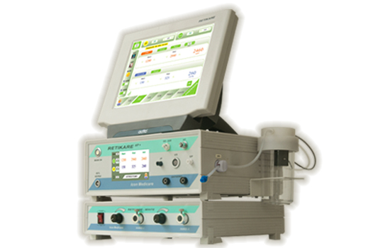
The Dual Linear control feature allows for simultaneous control of either flow or vacuum, and ultrasound power. Additionally, surgeons can use both flow and vacuum response in the same procedure. The powerful Quad-crystal ultrasound handpiece delivers excellent cutting with smooth efficiency. Programming options allow for virtually unlimited parameters of storage and surgeon-controlled mode switching. With its modular design, the Protege system integrates innovation for today and the future.
FEATURES
- Posterior Vitrectomy.
- 3000 CPM / 7500 CPM.
- 500 mmHg Venturi Aspiration.
- Independent Dual Led Light Source.
- 23/25g Capability.
- SOI/SOR & GF/IP.
- Linear/Fixed/Dual Settings.
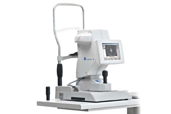
ZEISS IOL MASTER 500
The IOLMaster® 500 from ZEISS is the gold standard in optical biometry, with more than 100 million successful IOL power calculations to date. The ZEISS IOLMaster 500 is a great choice for cataract surgeons looking for a reliable, fast and easy-to-use optical biometer for measurements they can depend on.
ZEISS OCT STRATUS
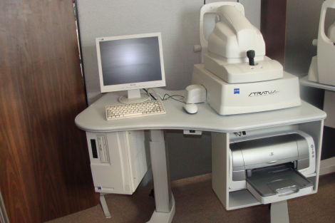
The ZEISS Stratus OCT Model 3000 (Stratus OCT) enables examination of the posterior pole of the eye at an extremely fine spatial scale, without surgical biopsy or even any contact with the eye.
The name Stratus OCT (derived from “stratum,” Latin for “layer”) refers to its unique ability of direct cross-sectional imaging of the layers of the retina. The Stratus OCT minimizes patient discomfort as it permits detailed examination of the retina and optic nerve head at the office or clinic. The Stratus OCT facilitates diagnosis and management of retinal diseases and glaucoma.
INTENDED USE
The Stratus OCT is intended for use as a diagnostic device to aid in the management of ocular diseases.
INDICATIONS FOR USE
The Stratus OCT is a non-contact, high resolution tomographic and biomicroscopic imaging device. It is indicated for in vivo viewing and axial cross-sectional imaging and measurement of posterior ocular structures, including retina, retinal nerve fiber layer, macula, and optic disc. It is intended for use as a diagnostic device to aid in the detection and management of ocular diseases including but not limited to macular holes, cystoid macular edema, diabetic retinopathy, age-related macular degeneration and glaucoma.
STRATUS OCT SYSTEM DESCRIBED
The Stratus OCT is a computer-assisted precision optical instrument that generates cross sectional images (tomograms) of the retina with ≤ 10 micrometers axial resolution. It works by using an optical measurement technique known as low-coherence interferometry.
STRATUS OCT DETIALS
The Stratus OCT incorporates optical coherence tomography technology to provide comprehensive imaging and measurement of glaucoma and retinal disease. Stratus OCT is the gold standard in vivo imaging device for the posterior segment and offers proven reproducibility for disease management.
The Stratus OCT provides real-time cross-sectional images and quantitative analysis of retinal features to optimize the diagnosis and monitoring of retinal disease and for enhanced pre- and post-therapy assessment. The device is beneficial for evaluation of cataract patients, pre- and post-operatively and for the assessment of early signs of glaucoma and glaucomatous change.
RETINAL NERVE FIBER LAYER (RNFL) ANALYSIS
Circular scans around the optic nerve head capture RNFL measurement of the peripapillary region. Analyses provide comparison of measurements to a normative database, demonstration of asymmetry and serial analysis.
OPTIC NERVE HEAD ANALYSIS
Radial line scans through the optic disc create cross-sectional and topographical data. The key analysis parameters include cup-to-disc ratios and horizontal integrated rim volume.
MACULAR THICKNESS ANALYSIS
Radial line scans provide cross-sectional images and analyses revealing retinal layers and macular condition. The change analysis feature illustrates change over time.
ZEISS FUNDUS CAMERA VISUCAM 500
The IOLMaster® 500 from ZEISS is the gold standard in optical biometry, with more than 100 million successful IOL power calculations to date. The ZEISS IOLMaster 500 is a great choice for cataract surgeons looking for a reliable, fast and easy-to-use optical biometer for measurements they can depend on.
VISUCAM® 500 features legendary ZEISS optics and non-mydriatic color fundus photography enabling you to photograph through pupils as small as 3.3mm. Superior patient comfort, more efficient workflow and improved eye care. The advantages of the comprehensive fundus platform VISUCAM 500 from ZEISS are obvious. The high-quality system provides everything you need for detailed diagnosis of typical eye diseases like diabetic retinopathy, glaucoma and AMD in a single workstation.
Advanced features such as fundus autofluorescence, easy stereo image handling and innovative assessment of macular pigment optical denstity (MPOD) are combined with intelligent auto functions that enable reproducible and intuitive imaging for every single patient eye.


ZEISS HFA3 830
NEW HUMPHREY FIELD ANALYZER – HFA3 VISUAL FIELD SERIES
HFA3 provides a streamlined and faster workflow with an array of remarkable new features designed to:
- Reduce visual field testing time with NEW SITA™ Faster.
- Mixed Guided Progression Analysis™ (GPA™): Inter-mixing of SITA Standard, SITA Fast, and SITA Faster.
- Reduce setup time with a single trial lens. Using liquid pressure, the new Liquid Trial Lens™ instantly delivers each patient’s refractive correction with the touch of a button.
- Improve confidence in test results with Rel EYE™**. Instantly review the patient’s eye position, at any stimulus point.
- Simplify operation with the intuitive new Smart Touch™ interface that novice users will appreciate.
- Gain peace of mind with seamless transferability of legacy data from the HFA II and HFA II-i to the HFA3.
- SITA Faster test is about half the time of SITA Standard and 70% of SITA Fast with the same reproducibility as SITA Fast. This may improve patient satisfaction with perimetric testing and reduce patient fatigue.
- Mixed GPA allows free mixing of SITA Faster, SITA Fast and SITA Standard and allows full access to the patient’s progression analysis including all SITA tests.
SIRIUS MASTER
Combines placido disk topography with Scheimpflug tomography of the anterior segment. Sirius provides information on pachymetry, elevation, curvature and dioptric power of both corneal surfaces over a diameter of 12 mm. All biometric measurements of the anterior chamber are calculated using 25 sections from the cornea. Measurement speed reduces the effect of eye movement producing a high quality accurate measurement. In addition to the clinical diagnosis of the anterior segment the most common uses are refractive and cataract surgery where an IOL calculation module is available. Objective examinations provide an accurate measurement of pupil diameter in scotopic, mesopic and photopic conditions, when combined with the corneal map they can be used for refractive surgery planning and follow-up.
ATLAS 9000 CORNEAL TOPOGRAPHY SYSTEM
- Simply Accurate for Maximum Productivity.
- Proven Placido Disk Technology with patented Cone-of-Focus™ Alignment System.

- SmartCapture™ Image Analysis Technology analyzes multiple images during alignment and automatically selects the highest quality image.
- MasterFit™ II Contact Lens Software helps streamline the fitting of gas permeable (GP) lenses and guides you through difficult and speciality fits.
- Data compatibility with previous generation ATLAS Corneal Topography Systems to facilitate data management and patient follow up.
GREEN SCAN LASER PHOTOCOAGULATOR
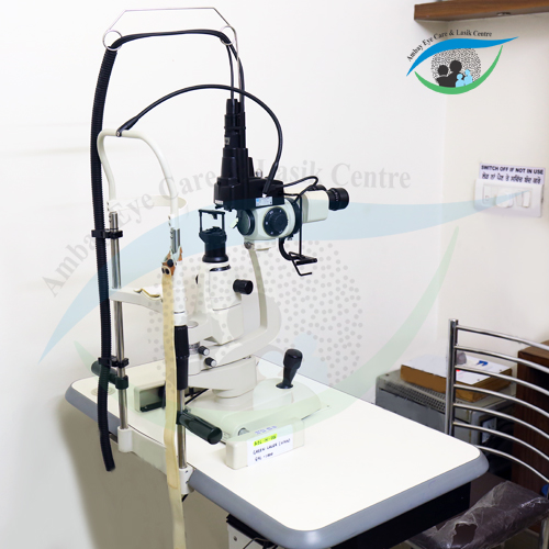
THE SMALL, INCREDIBLY VERSATILE GREEN LASER PHOTOCOAGULATOR
The GYC-500 Vixi / GYC-500 is a solid state green laser that achieves stable treatment outcomes for multiple applications including, retinal photocoagulation, trabeculoplasty and iridotomy.
The user-friendly features include a compact and lightweight design, and a wide range of delivery options allowing versatility for in-office use and the surgical suite.
LIGHTWEIGHT AND COMPACT DESIGN
This multifunction laser is housed in a small console. The space-saving design allows portability to virtually any room. 5.7-inch Color LCD with Touchscreen Control Box.
An intuitive graphic user interface and easy-to-read touch screen color LCD allows quick and easy setup and verification of the scan pattern and treatment parameters.
POP-UP WINDOW
The pop-up window appears once the displayed value, such as POWER, TIME, and INT is selected. The surgeon can easily make changes to these laser values.
STORED PHOTOCOAGULATION DATA
For flexibility in treating different types of clinical cases, 10 sets of photocoagulation data (power output, emission time, interval time, scan pattern and spacing) can be stored. Each set can be quickly retrieved with one-touch operation.
REGISTRATION OF CONTACT LENS MAGNIFICATION
Up to 5 contact lens magnifications can be registered. Confirmation of actual spot size on the retinal surface is easily performed by selecting the registered contact lens.
TREATMENT SUMMARY
Photocoagulation data can be displayed in one screen for review and output in XML format for saving the treatment.
HIGH RELIABILITY
A digitally controlled instant duty cycle permits the laser to be used at very fast speeds and high powers for extended periods of time without failure. The GYC-500 provides many years of superior, reliable performance.
STABLE AND RELIABLE GREEN LASER
The GYC-500 Vixi / GYC-500 ensures stable laser output by using a solid state laser. Two cooling fans in the console maintain the correct internal temperature. The maximum room temperature during use is 35ºC (95ºF) which is within the range to treat retinopathy of prematurity cases which requires ambient room to be approximately 30ºC (86ºF).
SOLIC (SAFETY OPTICS WITH LOW IMPACT ON CORNEA)
The SOLIC optical design is incorporated into all delivery units, ensuring low energy density on the cornea and lens, even for large spot sizes.
SCAN PATTERN OPTIONS
Incorporating Vixi, scan delivery units, into the GYC-500 enables laser treatments with various scan patterns. The GYC-500 Vixi enhances treatment efficiency and reduces patient chair time.
MULTIPLE SCAN PATTERNS
The GYC-500 Vixi has 22 preprogrammed scan patterns to allow treatment of various retinal pathologies.
AUTO FORWARD
Once photocoagulation is completed in one region, the GYC-500 Vixi allows automated advancement to the next region for delivery of the next scan pattern during photocoagulation. This feature allows the surgeon to concentrate on focus adjustment.
NIDEK YAG LASER 1800
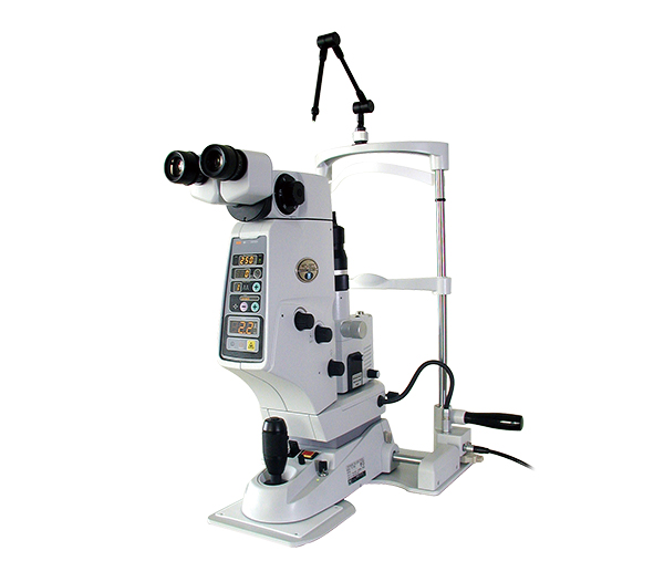
NIDEK’s compact YC-1800 ophthalmic photodisruptor offers the latest in innovative laser delivery and output technologies. Fast operation and ultra adjustability make the YC-1800 YAG Laser treatment system the finest available anywhere.
KEY FEATURES:
- High-res optics for exact laser-treatment location.
- S-Switch allows easy change of parameters while holding the joystick.
- Easily upgraded to the YAG/Green Combo system.
PORTABLE & USER-FRIENDLY DESIGN
IMPROVED OPERABILITY
The “S-Switch” located on the joystick provides high operability, allowing doctors to change parameters (Energy up, Energy down and Ready / Standby*) while holding the joystick. Permits faster and easier operation, and eliminates need to look away from oculars to make parameter adjustments.
ONE-TOUCH LOCK
The YC-1800 can smoothly slide back and forth and around, and the unit can be easily fixed and released at anywhere you like with the one-touch lock, offering high operability with improved safety.
COMPACT & SLIM DESIGN
The YC-1800 is Nidek’s smallest and lightest ophthalmic photodisruptor available and can be easily transported. Compact and slim design also allows greater flexibility in locating your arm rest.
RELIABILITY AND SAFETY
RELIABLE LASER OUTPUT
The YC-1800 employs the new technology to control the pulse number under the CPU “D-Pulse”, providing higher stability against environmental conditions and change over time.
FAST OPERATION
High-speed 3Hz firing rate offers very practical operation when encountering a moving eye or other patient difficulties. The YC-1800 can treat a wide variety of diseases, and its speed and efficiency allows comfortable operation.
GREAT NUMBER OF ENERGY SETTINGS
The YC-1800 offers 0.3-10mJ, continuously adjustable in increments of 0.1mJ, allowing the most precise tissue effect.
SUPER ADJUSTABLE ND : YAG OFFSET
- The YC-1800 can adjust the offset ±500 microns (25 micron steps) to meet the varied clinical needs.
- A different offset can be used for PMMA, silicone or acrylic lenses, and the offset can even be adjusted on the same IOL to compensate for a parallax effect in the periphery. This eliminates the need to manually defocus, permits a clear field of view, and minimizes lens pitting.
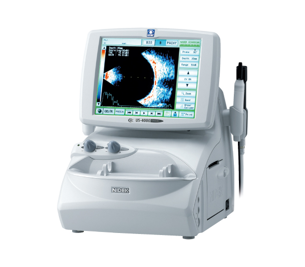
NIDEK B SCAN US-4000
FEATURES :
- Three-in-One unit of B-scan, Biometer, and Pachymeter.
No PC required. - Internal printer for instant printout of B-scan image.
- Tiltable 8.4-inch XGA color LCD for user-friendly operability.
USB and LAN interfaces for easy data storage.
B MODE
Scanning 400 lines over 60° provides high quality images, which are essential for accurate analysis.
BIOMETRY
Using new algorithms, axial length measurements and IOL power calculations are performed twice as rapidly as the conventional model.
PACHYMETRY
The pachymetry mode offers precise measurement of the corneal thickness within a ±5 μm error.
NIDEK ARK 510A
The ARK-510A provide exceptional measurement accuracy and highly tuned, dependable performance by combining the innovative measuring principle “Pupil Zone Imaging Method” and unique technology “SLD (Super Luminescent Diode).” The Pupil Zone Imaging Method is designed for refraction measurement by analyzing a wider area (maximum 4mm diameter) to obtain more reliable and realistic subjective refraction data. SLD combines a highly sensitive CCD for enhanced image quality and provides sharper, more vivid images than LED. Measurement capability is greatly improved even in eyes with certain opacification such as dense cataracts and IOL implanted eyes. The ARK-510A offers incomparable accuracy and consistency in keratometry measurement through use of a mire ring for enhanced alignment and observation.
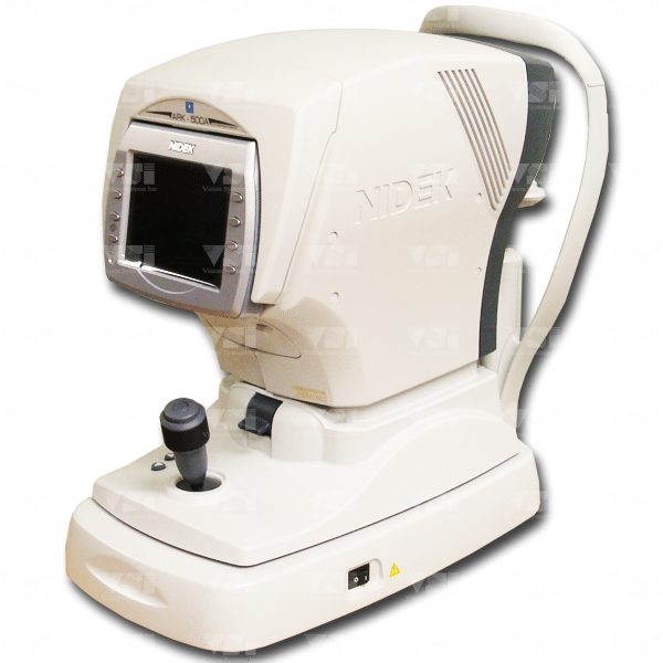

NIDEK AR-1 AUTOREFRACTOR
The AR(K)-1 ensures accurate refraction measurement (and keratometry). The large pupil zone imaging method enables the measurement of wide area refraction of up to 6 mm diameter. Fogging is performed after correcting the patient’s astigmatism to minimize the interference with accommodation even in high astigmatism. The SLD/CCD combining system improves measurement capability even in dense cataractous eyes.
NIDEK NCT NT-530P
FEATURES :
- Enhanced combination unit of non contact tonometer and pachymeter.
- Automatic calculation of compensated IOP.
- Advanced APC (Auto Puff Control) and noise reduction for comfortable tonometry measurement.
- Tiltable 5.7-inch color LCD for user-friendly operability.
3-D auto tracking, auto shot, and auto complete. - ACA mode.
DETAILED INFORMATION AUTOMATIC CALCULATION OF COMPENSATED IOP
The IOP compensation is automatically calculated with automatically measured patient’s central corneal thickness.
APC (AUTO PUFF CONTROL)
The APC achieves a quieter and softer air puff for patient’s more comfort. Tiltable 5.7-inch color LCD The 5.7-inch color LCD with tilting function offers easy operation even for a standing operator.
3-D AUTO TRACKING, AUTO SHOT, AND AUTO COMPLETE ACA MODE
The ACA mode allows the operator to capture an image of the anterior chamber angle with the Scheimpflug image.


NIDEK NT-510
FEATURES :
- IOP correction by corneal thickness.
- Advanced APC (Auto Puff Control) and noise reduction for comfortable tonometry measurement.
- Tiltable 5.7-inch color LCD for user-friendly operability.
- 3-D auto tracking, auto shot, and auto complete.
APC (AUTO PUFF CONTROL)
The APC provides a quieter and softer air puff for patient’s more comfort.
TILTABLE 5.7-INCH COLOR LCD
The 5.7-inch color LCD with tilting function offers easy operation even for a standing operator.
3-D AUTO TRACKING AUTO SHOT AUTO COMPLETE
SONOMED 100A+ A-SCAN
The Sonomed 100A+ Microscan A-scan offers a portable, affordable, and compact A-scan, with extreme accuracy, repeatable measurements and reliability. The combination of a high frequency, low noise probe and fast precise algorithm enables scan capture immediately upon application of the probe along the visual axis.
Axial length, ACD, and lens thickness are provided for each scan. Group up to five scans and the Micorscan 100A+ automatically calculates average axial length and standard deviation. Easily review each scan, delete outlying scans, and add new scans, as desired. Choose from one of four IOL formulas.

SONOMED 300A+ A-SCAN
The PacScan Plus 300A+ offers a portable, digital A-scan , with a large color touch screen, on-board memory and USB interface for EMR archiving, extreme accuracy, repeatable measurements and reliability. The combination of a high frequency, low noise probe and fast precise algorithm enables scan capture immediately upon application of the probe along the visual axis.
The PascScan Plus A-scan offers built-in immersion capabilities and up to nine IOL formulas, including three post-refractive formulas. Axial length, ACD, and lens thickness are provided for each scan. Group up to five scans with average axial length and standard deviation automatically calculated. Easily review each scan, delete outlying scans, and add new scans, as desired. Customizable tissue velocities of each structure and highly-developed automatic scan recognition algorithms ensure accurate and repeatable measures. Built-in calibration check ensures continued accuracy of system. Large color touch screen operation Complete measurement and calculation record within seconds Ability to store up to five different user profiles Portable, compact, and lightweight On-board memory storage and USB interface Built-in printer and probe storage.
TOPCON OPHTHALMOMETER OM-4
A professional for precise objective measurements of the corneal radius of curvature plus accurate measurements of the radius of curvature of contact lenses compactly-designed and very easy to use by one hand operation.


ZEISS SLITLAMP SL-130
The SL 130 is the instrument of choice for eye care professionals requiring a slit lamp with premium optical quality and high-precision mechanics to perform the complete range of eye care procedures.
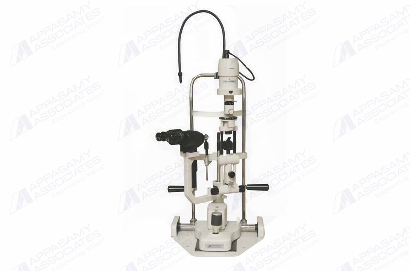
APPASAMY SLITLAMP SL-AIA 11
Features:
- Stereo Microscope Type: Galilean
- Magnification Changer: Drum rotation
- Working Distance: 100 mm
- Objective Lens Focal Length: f 100 mm
- Eye Pieces (Large Optics): 12.5x
- Total Magnifications: 10x, 16x, 25x
- Real Field of View: 27, 16, 11 mm
- Inter Pupillary Distance: 55 – 75 mm
- Diopter Adjustment Range: -6 D to +6 D
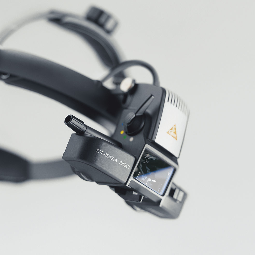
HEINE OMEGA®500 BINOCULAR INDIRECT OPHTHALMOSCOPE
Unique “Synchronized Adjustment of Convergence and Parallax” for high quality, stereoscopic fundus images through any pupil size. Precise selection of the observation and illumination optics for small pupils down to 1.0 mm. Excellent optical performance due to the multi-coated illumination system. Exact vertical alignment of the illumination with the observation path further minimising the reflections. Due to the mounting of the optics on an aluminum frame, the OMEGA500 is solid, long lasting, and is guaranteed to be dustproof. The HC50 L Headband Rheostat controls the LED illumination as well as the XHL Xenon-Halogen illumination.
PHILIPS CARDIAC MONITOR G30
- 10.4″ color TFT display.
- Maximum 8 waveforms display.
- 7-Lead ECG simultaneous display.
- ST and arrhythmia analysis.
- Optional dual-channel IBP and CO2 monitoring.
- Drug dose calculation and titration table.
- OxyCRG dynamic view display.
- Applications from neonate to adult.
- LAN networking capability to UT4800 Central Monitoring System.
- External VGA interface

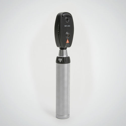
HEINE DIRECT OPHTHALMOSCOPE

